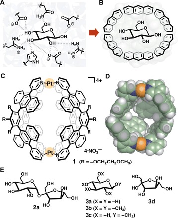
Fig. 1. Cartoon representation of saccharide recognitions and structures of a polyaromatic nanocapsule and saccharides.

Recognition of d-glucose (A) in a hydrogen-bonding cavity modeled after the binding site of sucrose hydrolase (E322Q-glucose complex) from Xanthomonas axonopodis pv. glycines (6) and (B) in a polyaromatic cavity. (C) Coordination-driven polyaromatic nanocapsule 1 and (D) its slice through the center of the crystal structure [space-filling model; substituents (R) are replaced by hydrogen atoms for clarity]. (E) d-Sucrose (2a), glucose derivatives 3a to 3c, and d-fructose (3d) used as guest molecules.
