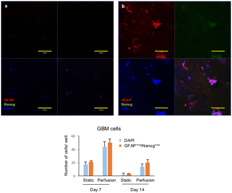
Figure 8.
GBM microtumours embedded in Matrigel, formed by primary cells isolated from a clinical sample. (a) Static culture of GBM microtumours. (b) Perfusion culture of GBM microtumours. GBM microtumours were formed by embedding primary cells in Matrigel, and then the 3D structures were extracted as described in Materials and Methods, followed by staining with astrocyte differentiation marker glial fibrillary acidic protein (GFAP, red fluorescence) and pluripotency marker Nanog (green fluorescence). Scale bar: 100 µm. Quantification of fluorescent cells shown in bar graph.

