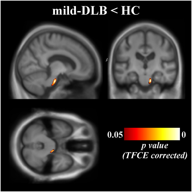
Figure 3.

Patterns of significant white matter loss in mild-DLB patients mild-DLB, dementia with Lewy bodies at the stage of dementia; HC, healthy controls. Results are expressed as p-value from permutation tests with threshold-free cluster enhancement corrected for multiple comparisons, and are superimposed on the mean MNI-standardized MRI T1-weighted image of the patients at the stage of dementia and healthy controls. Left is left side of the brain. Coordinates of the multiplanar image in the MNI space: x = 11; y = −18; z = −30.
