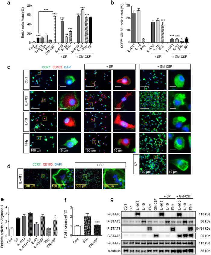
Figure 4.
SP is a mitogen for macrophages and affects M2 polarization in a dominant manner even in the presence of the Th1 cytokine INFγ. (a) BrdU incorporation analysis revealed that SP was less potent at inducing proliferation than GM-CSF and that SP and GM-CSF had a synergistic effect on proliferation on all types of macrophages at 3 d. Fold change of BrdU incorporation in MΦGM-CSF was shown in Supplementary Figure 2a. The number of BrdU+ cells was counted (5 random fields/coverslip) at 100 × magnification (0.85 mm2) (n = 3). (b–c) The percentage of CCR7loCD163+ M2 macrophages was maintained in M2aIL-4/13 and M2cIL-10 subtypes in the presence of SP but completely diminished after 3 d of GM-CSF treatment. SP-induced M2 macrophages in M1INFγ showed similar proportions of the M2aIL-4/13 and M2cIL-10 subtypes (Green = CCR7, Red = CD163, Blue = DAPI). The number of CCR7loCD163+ M2 cells was counted (5 random fields/coverslip) at 100 × magnification (0.85 mm2) (n = 3). (d) IL-4/13-induced MGCs were still present in response to SP co-treatment at 3 d (Green = CCR7, Red = CD163, Blue = DAPI) (n = 6). (e) The activity of Arginase-1 was increased at 6 h in response to IL-4/13, IL-10 or IFNγ co-treatment with SP, as determined by Arginase-1 activity assay. SP-induced M2 polarization is dominantly working over Th1 cytokines, IFNγ (n = 6). (f) The iNOS activity assay revealed a decrease in IFNγ activity after SP co-treatment compared with that after IFNγ treatment at 1 d (n = 3). (g) SP did not activate STAT1, STAT3, STAT5, or STAT6, and SP co-treatment did not affect specific cytokine-mediated STAT expression such as STAT6 by IL-4/13, STAT3 by IL-10, STAT1 by IFNγ, and STAT5 by GM-CSF at 6 h. The western blot samples derived from the same experiment and full-length blots are presented in Supplementary Figure 23. (n = 2). Data represent the mean ± SEM (*p < 0.05, ***p < 0.001, unpaired t test). Scale bar = 500 μm (d), 100 μm (c, high-magnification image inset in d) and 10 μm (high-magnification image inset in c).

