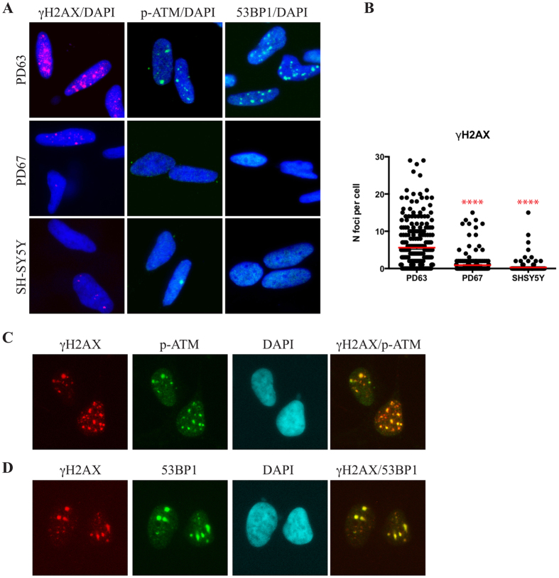
Figure 3.
DNA repair foci accumulate in differentiated PD63 cybrids. (A) Immunolocalization of γH2AX (red), ATMpS1981 (p-ATM) (green) and 53BP1 (green) in PD63, PD67 and SH-SY5Y cells after differentiation. Cells were seeded and differentiated on coverslips and immunostained with specific antibodies. (B) The number of foci per nucleus in the three cell lines was quantified with CellProfiler as described in Material and Methods. Red lines indicate the means of γH2AX foci per nucleus. For these analyses more than 200 cells for each condition were analysed; **** = p < 0.0001. (C) Immunofluorescence showing the colocalisation of γH2AX (red) and ATMpS1981 (p-ATM) (green) in differentiated PD63 cybrid cells. (D) Immunofluorescence showing the co-localization of γH2AX (red) and 53BP1 (green) in differentiated PD63 cybrid cells. Nuclei were stained by DAPI.

