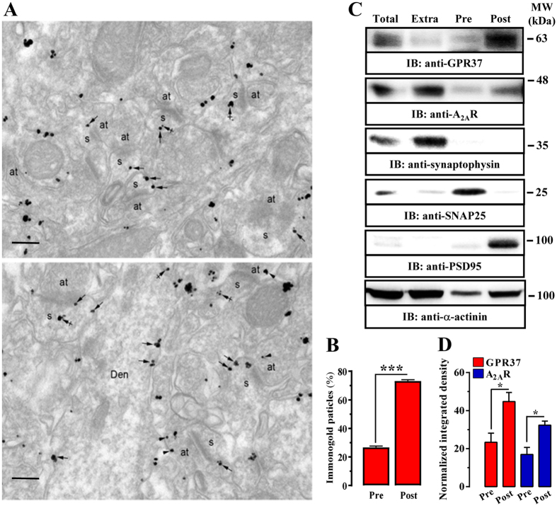
Figure 2.
Subsynaptic distribution of GPR37 in the mouse striatum. (A) Electron micrographs showing immunoparticles for GPR37 in the striatum of GPR37+/+ mice using the pre-embedding immunogold technique. GPR37 immunoparticles were abundant on the extrasynaptic plasma membrane (arrows) of dendritic spines (s) of striatal neurons contacted by axon terminals (at). Few immunoparticles were observed at intracellular sites (crossed arrows) in dendritic spines (s) (upper panel). Immunoparticles for GPR37 were also localized to the extrasynaptic plasma membrane (arrowheads) of axon terminals (at) establishing asymmetrical synapses with spines (s). In dendritic shafts (Den), immunoparticles for GPR37 were mainly found at the plasma membrane (arrows) (lower panel). Scale bars: 200 nm. (B) Bar graphs showing the percentage of GPR37 immunoparticles at post- and presynaptic compartments. The data are expressed as mean ± SEM of three independent experiments (***P < 0.001; Student’s t-test). (C) Representative immunoblots showing GPR37 and A2AR immunoreactivity in striatal synaptic fractions. Striatal synaptosomes (Total) were subcellularly fractionated (see Materials and Methods section) into extrasynaptic (Extra), presynaptic active zone (Pre) and postsynaptic density (Post) fractions, which were analyzed by SDS-PAGE (20 μg of protein/lane) and immunoblotted using rabbit anti-GPR37-N, goat anti-A2AR, rabbit anti-synaptophysin, mouse anti-PSD-95, mouse anti-SNAP-25 and rabbit anti-α-actinin antibodies. The primary antibodies were detected using a horseradish peroxidase (HRP)-conjugated goat anti-rabbit IgG, HRP-conjugated goat anti-mouse IgG, HRP-conjugated rabbit anti-goat IgG and chemiluminescence detection (see Materials and Methods). (D) Relative quantification of GPR37 enrichment in striatal presynaptic and postsynaptic fractions. The intensities of the immunoreactive bands on the immunoblotted membranes corresponding to extrasynaptic, presynaptic (Pre) and postsynaptic (Post) fractions were measured by densitometric scanning. Values were first normalized to the loading control (i.e. α-actinin) and then to the amount of GPR37 or A2AR in the total fraction. Data are expressed as mean ± SEM of three independent experiments (*P < 0.05, Student’s t-test).

