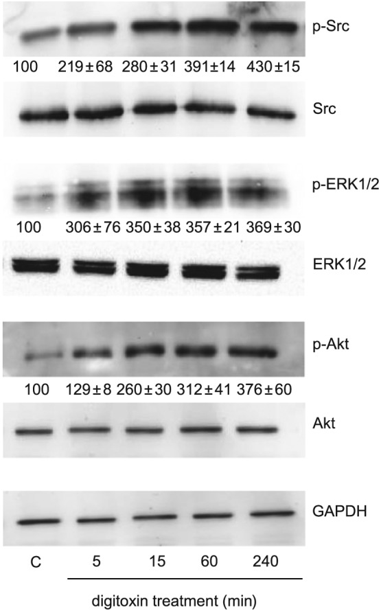
Figure 6.

Signalling pathways activated by digitoxin in HUVEC. Cells were grown in six‐well plates and, after reaching confluence, were incubated overnight in culture medium containing 1% FBS. Then cells were stimulated with digitoxin (10 nM) for the time indicated. Cell lysates were analysed by Western blotting. Representative blots of an experiment showing the expression of phosphorylated and total Src, phosphorylated and total ERK1/2, phosphorylated and total Akt; GAPDH expression was used as loading control. Below each blot of phosphorylated proteins is shown the densitometric analysis of the bands, normalized to GAPDH levels, expressed as % of control (mean ± SEM from three independent experiments).
