Figure 6.

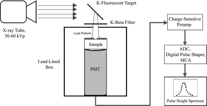
Schematic of the pulse height spectroscopy apparatus used in our experiments. Monoenergetic x‐rays produced via K‐fluorescence excited the sample, which was coupled to a PMT. PMT output was fed to a digital pulse height analysis module.

Schematic of the pulse height spectroscopy apparatus used in our experiments. Monoenergetic x‐rays produced via K‐fluorescence excited the sample, which was coupled to a PMT. PMT output was fed to a digital pulse height analysis module.