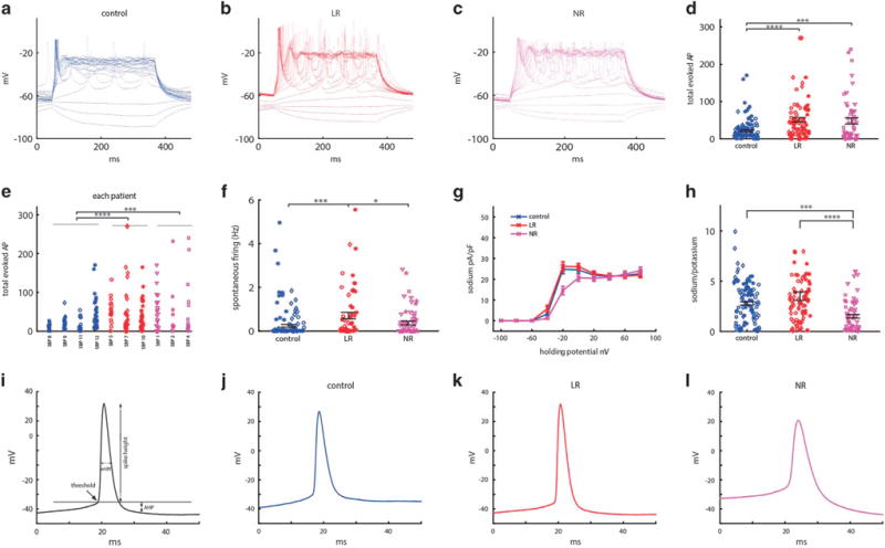
Figure 2.

Dentate gyrus granule-cell-like hippocampal neurons derived from patients with bipolar disorder (BD) are hyperexcitable at 3.5 weeks (20–30 days) postdifferentiation when comparing control patients’ neurons (control n = 95 cells from four lines), lithium-responsive (LR) patients (LR n = 84 cells from three lines) and lithium-non-responsive (NR) patients (NR n = 63 cells from three lines), and spike shape is different between the three groups (control, LR and NR). (a–c) Representative recordings of evoked action potentials in current clamp mode of a (a) control (b) LR and (c) NR neuron. (d) Total number of evoked action potentials in 35 depolarization steps shows hyperexcitability of BD neurons. (e) Patient-by-patient plot of the total evoked action potentials. (f) Spontaneous action potential rate measured when the cell is typically held between −45 and −50 mV for 60 s. (g) Average of normalized sodium currents as a function of membrane holding potential display lower sodium current in NR neurons. (h) Average ratio of sodium current measured at −20 mV and slow potassium current measured at 20 mV display lower ratios of the currents in NR neurons. (i) Demonstration of action potential features. (j–l) Representative trace of an action potential of a control (j), LR (k) and NR neuron (l) displays a different spike shape between the three groups (see Supplementary Figure 1 for averages of the different features). LR neurons have a higher amplitude and narrower action potential compared with control neurons (k), whereas NR neurons have a lower amplitude and broader action potentials, with a more depolarized threshold for evoking an action potential (l). Identical symbols indicate cells from the same cell line. *P<0.05, **P<0.01. ***P<0.001 and ****P<0.0001.
