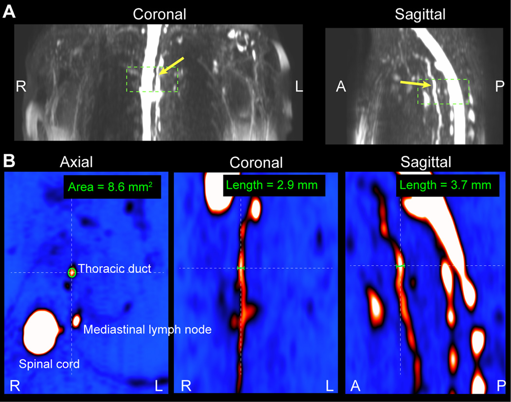
Figure 2.
Lymphatic vessel cross-sectional area measurement procedure. (A) Orthogonal representations of lymphangiography MIPs are generated to locate the lymphatic vessel of interest. This example is performed on the thoracic duct (yellow arrow) which is located anterior to the spinal cord. (B) The cross-sectional area is measured on magnified versions of the magnitude images themselves where vessel size can be confirmed in all three imaging planes.

