Figure 5.
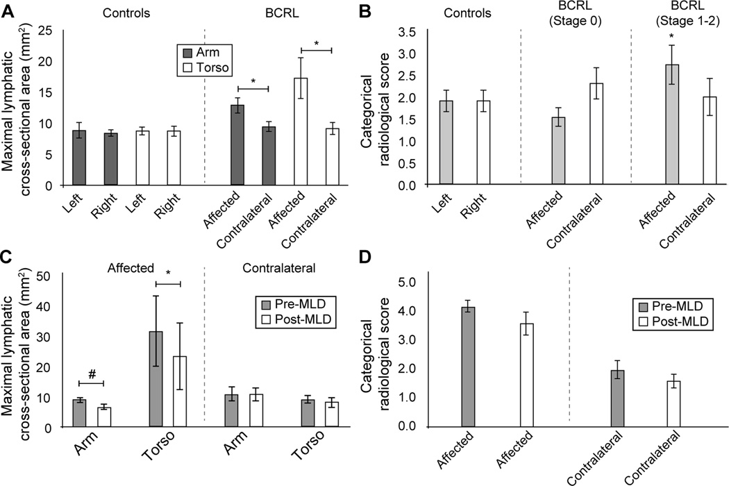
Summary of primary study findings. (A) Maximal lymphatic cross-sectional area did not differ significantly between left and right sides in controls, but was significantly elevated in the affected relative to contralateral side of patients. This was found both for arm and torso vessels and for both symptomatic (Stages 1–2) and sub-clinical patients. (B) The categorical scoring system was less sensitive to lateralizing disease. Neither left vs. right scores in controls, nor affected vs. contralateral scores in all patients, were significantly different. When only symptomatic patients were considered however, the scores in the affected side were significantly elevated relative to control scores. (C) In the subgroup of patients scanned before and after MLD, maximal lymphatic cross-sectional area was observed to reduce on the affected side only; this change was significant in the torso region and just beyond significance criteria in the arm region. (D) The categorical score changes pre- vs. post-MLD were not significant, however the effect sizes for the reductions were large on both the affected (Cohen’s d=0.81) and contralateral (Cohen’s d=0.63) side. * two-sided p<0.05; # one-sided p<0.05. Values summarized are mean ± one standard error of the mean. Standard deviations are summarized as well in Table 2.

