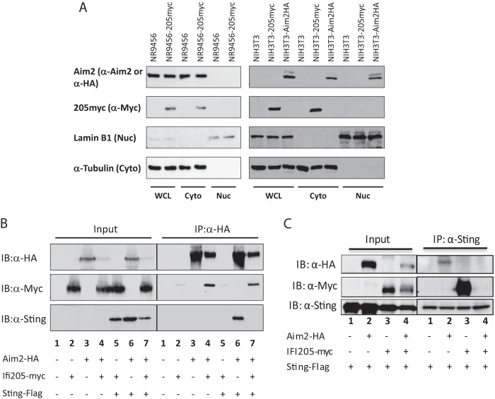
FIG 6 .
Interaction of IFI205, AIM2, and STING. (A) Intracellular localization of IFI205 and AIM2 in NR9456 and NIH 3T3 cells. Lysates of cells stably expressing IFI205myc or AIM2HA were fractionated into cytoplasmic and nuclear fractions and then subjected to Western blotting with the antibodies indicated. Lamin B1 and α-tubulin were used as markers for the nucleus and cytoplasm, respectively. A single experiment was done for each panel. (B, C) Co-IP of IFI205myc, AIM2HA, and STING-FLAG. HEK293T cells were transfected with tagged proteins as indicated. Cells were lysed 48 h after transfection and immunoprecipitated (IP) with anti-HA (B) or anti-FLAG (C) antibodies. Proteins were detected by Western blotting with the antibodies indicated. The co-IP was repeated three times and gave the same results. WCL, whole-cell lysate; Cyto, cytoplasm; Nuc, nucleus; IB, immunoblotting.

