Fig. 1.

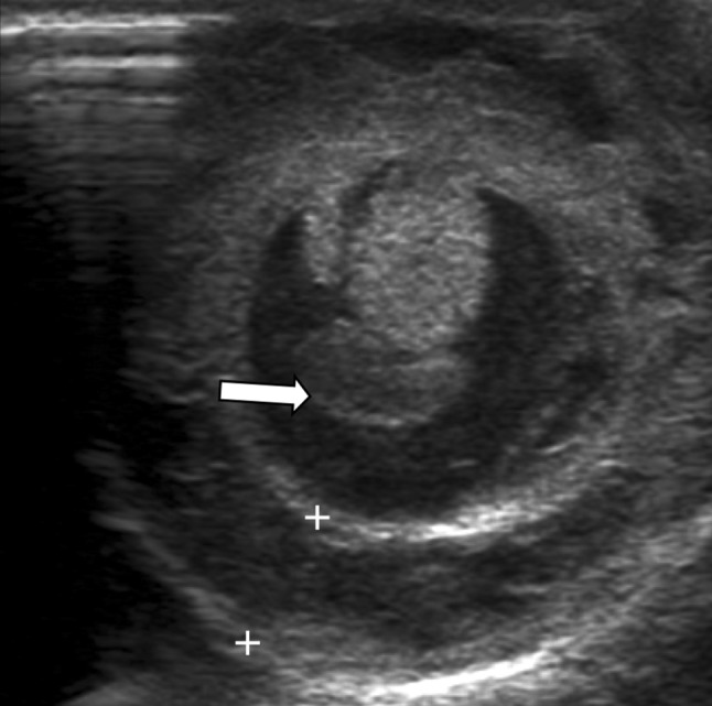
Transverse sonographic view of the right hemi-scrotum showing the normal testis and epididymal body surrounded by fluid. Adjacent omentum (arrow) is seen. Grossly edematous and thickened scrotal wall (within calipers) is also appreciated

Transverse sonographic view of the right hemi-scrotum showing the normal testis and epididymal body surrounded by fluid. Adjacent omentum (arrow) is seen. Grossly edematous and thickened scrotal wall (within calipers) is also appreciated