Fig. 2.

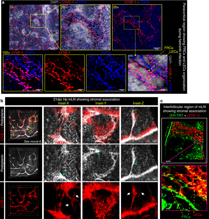
LECs and FRCs form intimate contacts in the area surrounding B-cell follicles. C57BL/6 J wild-type mice were infected with Hp and the entire chain of the mLN collected at 21 dpi. a 8 μm thick mLN cryosections showing combined immunofluorescence staining for FRCs (PDPN+; blue) and LECs (LYVE-1+; red) of Hp-infected mice are shown at various magnifications. Scale bar 50 μm. b Vibratome sections of ≥100 μm thick mLN showing 2D and 3D views of paracortical LECs (red; PDPN+LYVE-1+) and FRCs (gray; PDPN+LYVE-1−) lying in close vicinity of one another and surrounding a centrally located B-cell follicle. Insets (x, y, z) show higher magnification of FRCs and LECs. Green and white arrows highlight quiescent and sprouting lymphatic vessels respectively. c Vibratome sections of thick mLN section showing the mLN interfollicular region and highlighting associations between FRCs and LECs. All images are taken from a single mouse and are representative of two or more independent experiments, each including n ≥ 2 mice/group/time-point. See also Supplementary movie 6
