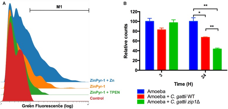
FIGURE 2.
Acanthamoeba castellanii cells reduce intracellular zinc levels in the presence of C. gattii. (A) Cytometry histogram of ZinPyr-1 fluorescence A. castellanii cells cultured in PYG (Control), PYG plus 10 μM zinc chelator TPEN (TPEN) and PYG plus 50 μM ZnCl2. (B) A. castellanii (1 × 10⁵ cells) and C. gattii WT or zip1Δ (1 × 10⁶ cells) were incubated at 1:10 ratio in PYG medium for 3 and 24 h at 30°C. The wells were washed with PBS and then incubated with Zinpyr-1 cell-permeable fluorescent probe for 30 min. After, washes with PBS were performed and the cells were collected for flow cytometry analysis. Data are shown as the mean ± SD from three experimental replicates per condition. The asterisks denote statistically significant differences between the conditions, as evaluated by Student’s t-test (∗p < 0.05; ∗∗p < 0.01).

