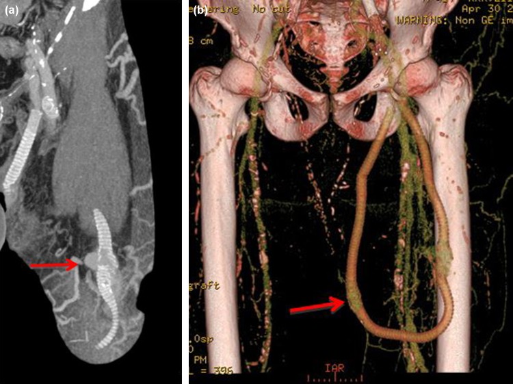
Figure 9.
(a) Maximum intensity projection (MIP) image of a left loop thigh AVG in a patient who presented with spontaneous bleeding over the AVG. It demonstrates localized breakdown of the graft material and pseudoaneurysm formation (arrow) associated with repetitive needling at the same site. This was urgently treated with insertion of a stent-graft. (b) 3D reconstruction of the same patient also shows early AVG breakdown in the medial limb of the graft (arrow).

