
Figure 6.
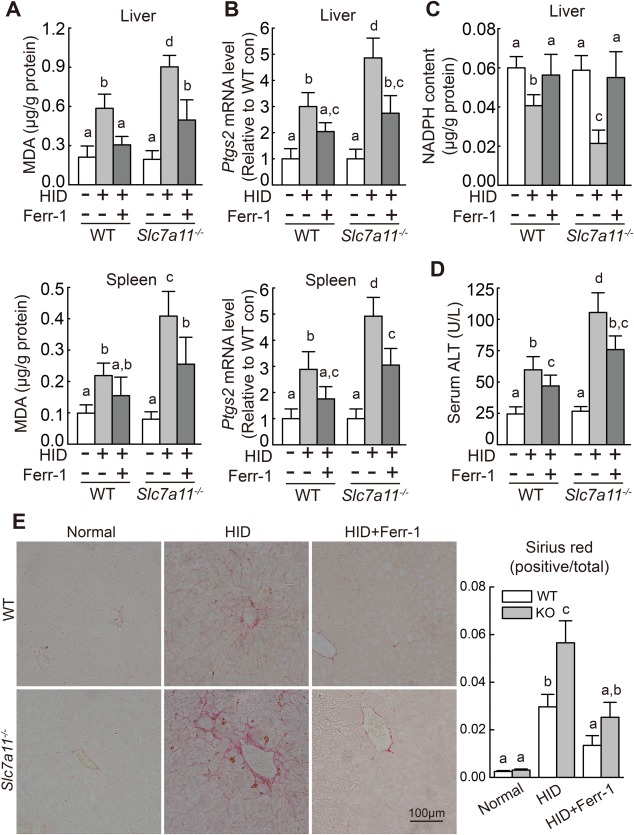
Iron‐overloaded Slc7a11–/– mice have increased levels of tissue ferroptosis. (A) Hepatic and splenic MDA content, (B) hepatic and splenic Ptgs2 mRNA, (C) hepatic NADPH content, and (D) serum ALT activity were measured in male wild‐type and Slc7a11–/– mice that were fed a normal‐iron diet or the HID with or without Ferr‐1 treatment (n = 6 mice/group). (E) (left) Liver sections were obtained from the indicated mice and stained with sirius red; (right) summary of staining intensity. mRNA levels in (B) were normalized to β‐actin mRNA and are expressed relative to the respective mean wild‐type control value. Significance was calculated using a one‐way ANOVA with Tukey's post hoc test, and groups labeled without a common letter were significantly different (P < 0.05). Abbreviations: con, control; KO, knockout; WT, wild type.