Figure 1.

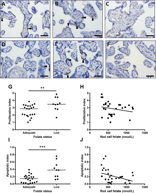
Effect of maternal folate status on placental cell turnover. Representative images of Ki67 and M30 immunohistochemistry in placentas from adolescents with (A and D) adequate (n = 24) and (B and E) low (n = 10) folate status. (C and F) Negative. Proliferation index (Ki67+ nuclei/total nuclei) was (G) elevated in placentas from women with low versus normal folate status (p < 0.01, Mann–Whitney test), and (H) inversely correlated to maternal RBC folate concentrations (r = –0.359, p < 0.05, Spearman's rank). Apoptotic index (M30+ cells/total nuclei) was (I) elevated in placentas from women with low versus normal folate status (p < 0.001, Mann–Whitney test), and (J) inversely related to maternal RBC folate concentrations (r = –0.426, p ≤ 0.01, Spearman's rank). **p < 0.01, ***p < 0.001. Scale bars = 50 μm. Arrowheads indicate positive staining.
