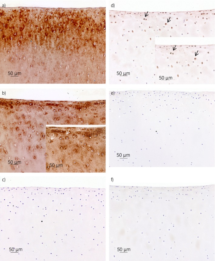
Figure 3.

Immunohistochemistry of cartilage explants stained with polyclonal antibodies against total cartilage oligomeric matrix protein a,b), against the cartilage oligomeric matrix protein fragment c,d) and negative (isotype) controls e,f). Explants a, c and e were unstimulated and kept in media for 25 days, and explants b, d and f were stimulated with interleukin‐1β (10 ng/mL) for 25 days. a) severe diffuse staining of the extracellular matrix (ECM); b) moderate cytoplasmic and pericellular staining (white arrows) and diffuse staining of the ECM (territorial and interterritorial); c) no staining; d) mild cytoplasmic and pericellular staining (black arrows); e,f) no staining. Scale bar = 50 μm.
