
Figure 4.
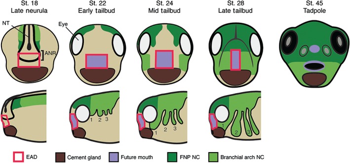
Relationship of the extreme anterior domain (EAD) and neural crest (NC) to the Xenopus mouth. Branchial arch (BA, light green) NC, and frontonasal prominence crest (FNP, dark green) delaminate from the dorsal neural tube (NT) at neurula. The EAD (red outline) is specified including cells arising from the anterior neural ridge (ANR, black outline) and cement gland (CG, brown) tissue. The EAD (purple) will contribute to the lining of the mouth (as well as the nostrils and anterior pituitary). FNP NC migrates anteriorly between the eyes to enter the face while first BA NC migrates bilaterally into the face (late neural‐mid tailbud). Subsequent to NC ingress, EAD ectoderm thins and lengthens to become the ‘pre‐mouth array’ (early‐mid tailbud). NC cells eventually differentiate to form the facial skeleton, including the palate, maxilla, mandible, and connective tissue.