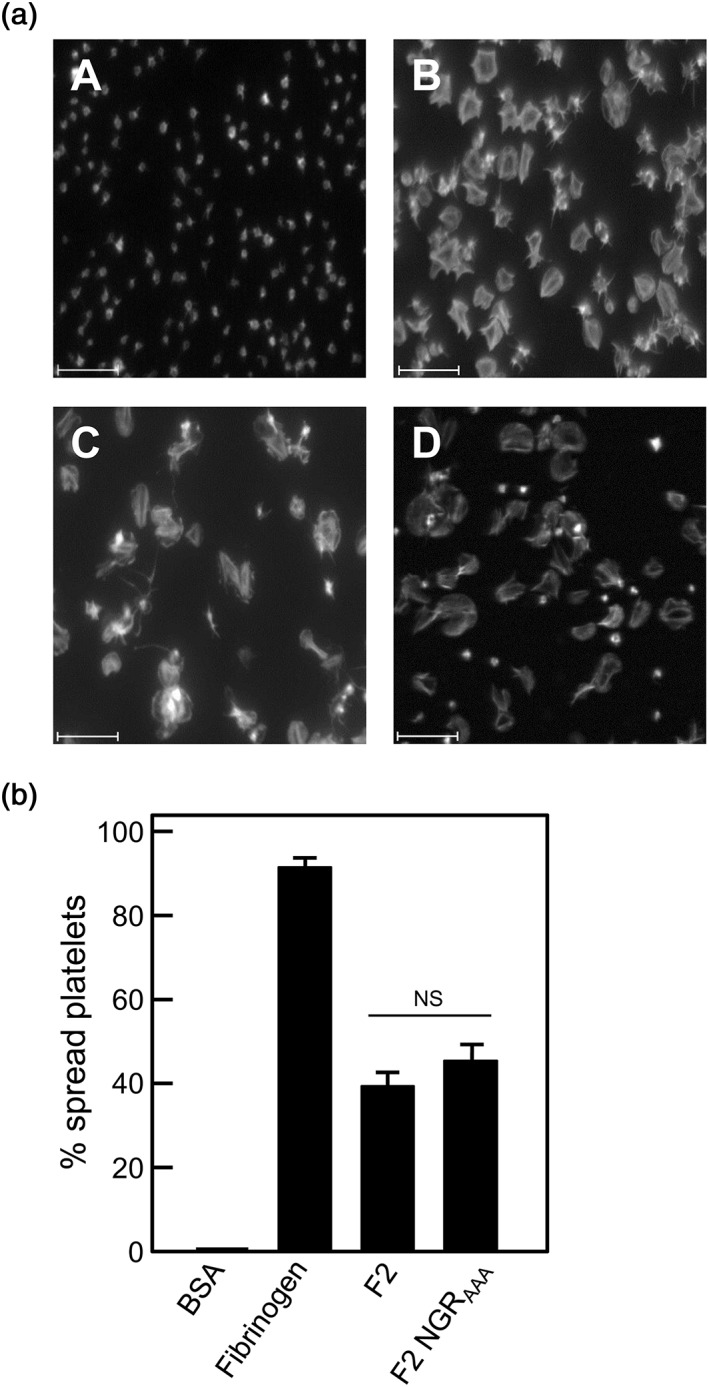
Figure 5.

Platelet spreading on immobilized recombinant PadA‐F2 region fragments under static conditions. (a) BSA (negative control), (b) fibrinogen (positive control), (c) recombinant PadA protein fragment F2, or (d) fragment F2 NGRAAA were immobilized onto microtitre plate wells (10 μg per well), and non‐specific binding sites were blocked with BSA. Gel‐filtered platelets were incubated with the immobilized substrates at 37°C for 45 min (2 × 10⁷ platelets per well), and platelet spreading was visualized by confocal microscopy. Scale bar = 15 μm. (b) Percentage of platelets spread on BSA (negative control), fibrinogen (positive control), and recombinant PadA protein F2 fragments immobilized onto glass slides. Error bars represent ±SEM from three independent experiments (n = 3). NS = not statistically significant
