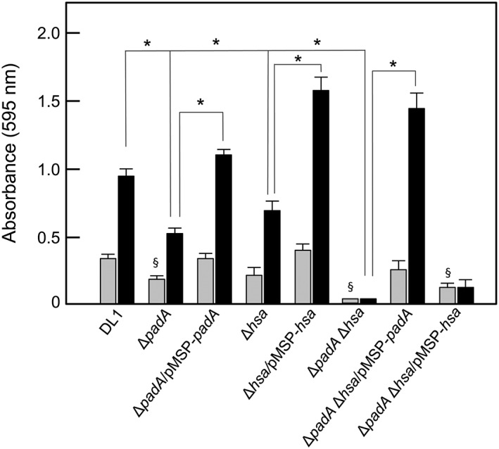
Figure 8.

S. gordonii strains adherence to salivary pellicle and biofilm formation. Cover slips were coated with salivary pellicle and incubated with streptococcal cells (5 × 10⁷ per well) for 2 hr at 37°C for adherence (grey columns) or in YPT‐Glc medium for 16 hr at 37°C for biofilm formation (black columns). Bacterial cells adhered, and biofilm biomass values were quantified by staining with crystal violet as described in 4. Expression of PadA or Hsa proteins by complemented strains was induced with 10 ng or 50 ng nisin ml−1, respectively. Error bars represent ±SEM from three independent experiments (n = 3). * P < 0.05 for biofilm comparisons indicated; § P < 0.05 for significantly different adherence versus DL1
