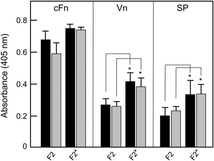
Figure 9.

Adhesion of PadA‐F2 region fragments to cellular fibronectin (cFn), vitronectin (Vn), or salivary pellicle (SP). cFn, Vn, or saliva were immobilized onto microtitre plate wells and non‐specific binding sites were blocked with BSA (black columns). Wells coated with cFn, Vn, or salivary glycoproteins were also incubated with 0.001 U neuraminidase (sialidase) for 2 hr at 37°C, washed, and blocked with BSA (grey columns). Recombinant proteins (100 μg ml−1) were incubated with substrata for 2 hr at 37°C and amounts bound measured with anti‐tetra‐His mouse antibodies and HRP‐conjugated anti‐mouse IgG antibodies as described in 4. F2, unmodified fragment; F2*, fragment containing NGR, RGT, and AGD motifs all alanine‐substituted to AAA. Error bars represent ±SEM from three independent experiments (n = 3). * P < 0.05 compared with corresponding F2 values
