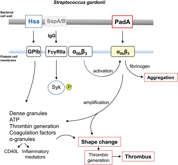
Figure 10.

Diagrammatic representation of some of the processes involved in platelet activation by S. gordonii. Cell wall‐anchored proteins Hsa and PadA interact with platelet membrane integrins GPIb and αIIbβ3 (GPIIbIIIa). Hsa captures platelets under flow (rolling) by binding GPIb, and possibly also αIIbβ3, and activates signalling cascades including FcγRIIa phosphorylation, leading to dense granule release (see Arman et al., 2014). PadA binds activated αIIbβ3, thus amplifying signals leading to shape change, thrombin production, coagulation, and thrombus formation. Platelet activation by S. gordonii can occur in the absence of specific IgG. However, with IgG present, there is evidence for activation (phosphorylation) of spleen tyrosine kinase (Syk‐P) through FcγRIIa. Conserved streptococcal surface protein antigens such as antigen I/II proteins (e.g., SspA/B) may also be involved in the overall process (Kerrigan et al., 2007). Physiologically, collagen activates Syk through GPVI, which is closely associated with FcγRIIa (not shown). Fibrinogen engages GPIIbIIIa (αIIbβ3), which also associates with FcγRIIa. CD40L (otherwise known as CD154) is up‐regulated in the platelet cell membrane and binds CD40+ cells such as endothelial cells and neutrophils, while soluble (released) CD40L further activates platelets
