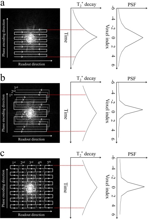
Figure 1.

(a) Single‐shot EPI trajectory samples k‐space very rapidly (20–40 msec per image). However, tissue decay causes signal loss at outer k‐space, which corresponds to high spatial frequencies, leading to image blurring. This blurring is quantified by the point spread function (PSF), which describes the extent of blurring of signal from nearby voxels (ie, a wider PSF corresponds to more blurring). Partial Fourier acquisition is illustrated here, which can effectively reduce the echo time and hence increase SNR. (b) Conventional segmented EPI (three segments shown here) and (c) readout segmented EPI (five segments shown here) significantly reduces the effective echo spacing (eg, from about 0.8 msec in SSH‐EPI to about 0.25 msec in segmented EPI and 0.32 msec in readout segmented EPI; parallel imaging is not considered here), leading to sharper PSF shapes. Note that we are only considering decay, but that T 2 decay will also be occurring. However, this effect does not in general change the PSF characteristics by much.
