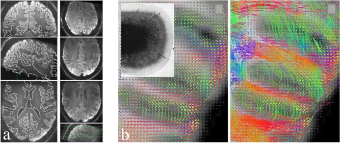
Figure 7.

High‐resolution (0.8 mm isotropic) diffusion MRI data acquired using a combination of reduced FOV and parallel imaging method at 7T. (a) Left column: Trace‐weighted images overlaid with white/gray matter boundaries obtained from an anatomical scan, demonstrating high geometric fidelity achieved by combining reduced FOV methods and parallel imaging. Right column: axial slices at different brain regions. (b) Fiber orientation density (left) and streamline tracking (right) based on the high‐resolution data, depicting white matter fiber tracts entering the cortex. Figure reproduced with permission from Ref. 66.
