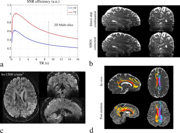
Figure 10.

(a) SNR efficiency plot for diffusion‐weighted spin‐echo sequence. Here, white matter is analyzed. For the T 1 value of white matter, the optimal TR is between 1–2 seconds, which is not compatible with full brain coverage using conventional 2D multislice acquisitions. Simultaneous multislice acquisition enables a short TR, but still faces limitations at high‐resolution scan. 3D multislab can achieve very short TR (2–3 sec) for full brain coverage, enabling a higher SNR efficiency. (b) Slab boundary artifacts in 3D multislab acquisition. First row shows the result of direct slab combination, where all slabs are concatenated with outermost slices discarded. Second row shows the correction using Nonlinear inversion for slab Profile ENcoding (NPEN) method, in which the artifacts are effectively reduced. (c) 1 mm isotropic resolution diffusion MRI data acquired at 7T using 3D multislab acquisition, demonstrating high SNR and good anatomical details. (d) Tractography of the cingulum bundle from in vivo data (top row) captures essentially the full extent of the cingulum bundle, including temporal and frontal lobe tracts and cortical projections into cingulate cortex. Tractography on postmortem data (from a different study) is shown as gold standard (bottom row).
