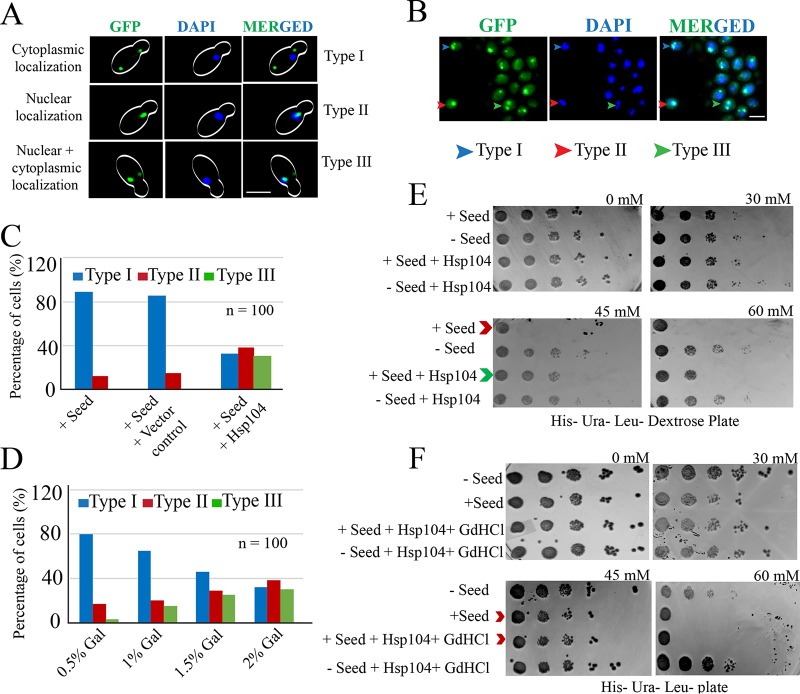
FIG 11.
Rescue of p53 amyloid due to overexpression of Hsp104. (A) Live-cell imaging to visualize p53-GFP signal in cells overexpressing Hsp104. Three types of cells were observed: type I, where cytoplasmic signal of p53-GFP was observed, suggesting localization of p53 aggregates in the cytoplasm; type II, where nuclear signal of p53-GFP was observed, suggesting functional nonaggregating p53 enters the nucleus; and type III, where both cytoplasmic and nuclear signals were observed, suggesting a fraction of p53 aggregates were rescued and became functional due to Hsp104 overexpression. Scale bar, 5 μm. (B) Field view showing all three types of cells. Scale bar, 3 μm. (C) Percentage distributions of the three types of cells with or without Hsp104. (D) Dose-dependent curing of p53 prion by Hsp104. (E) Growth defect observed due to inactivation of transcription of the HIS3 reporter for the cells harboring seeds. However, the defect was partially rescued when the same cells overexpressed Hsp104 (compare the rows indicated by the red and green arrowheads). (F) The growth rescue effect of Hsp104 in panel E was abolished upon treatment with GdHCl, and the growth of the treated cells became similar to the that of the plus-seed cells (compare the rows indicated by the red arrowheads).

