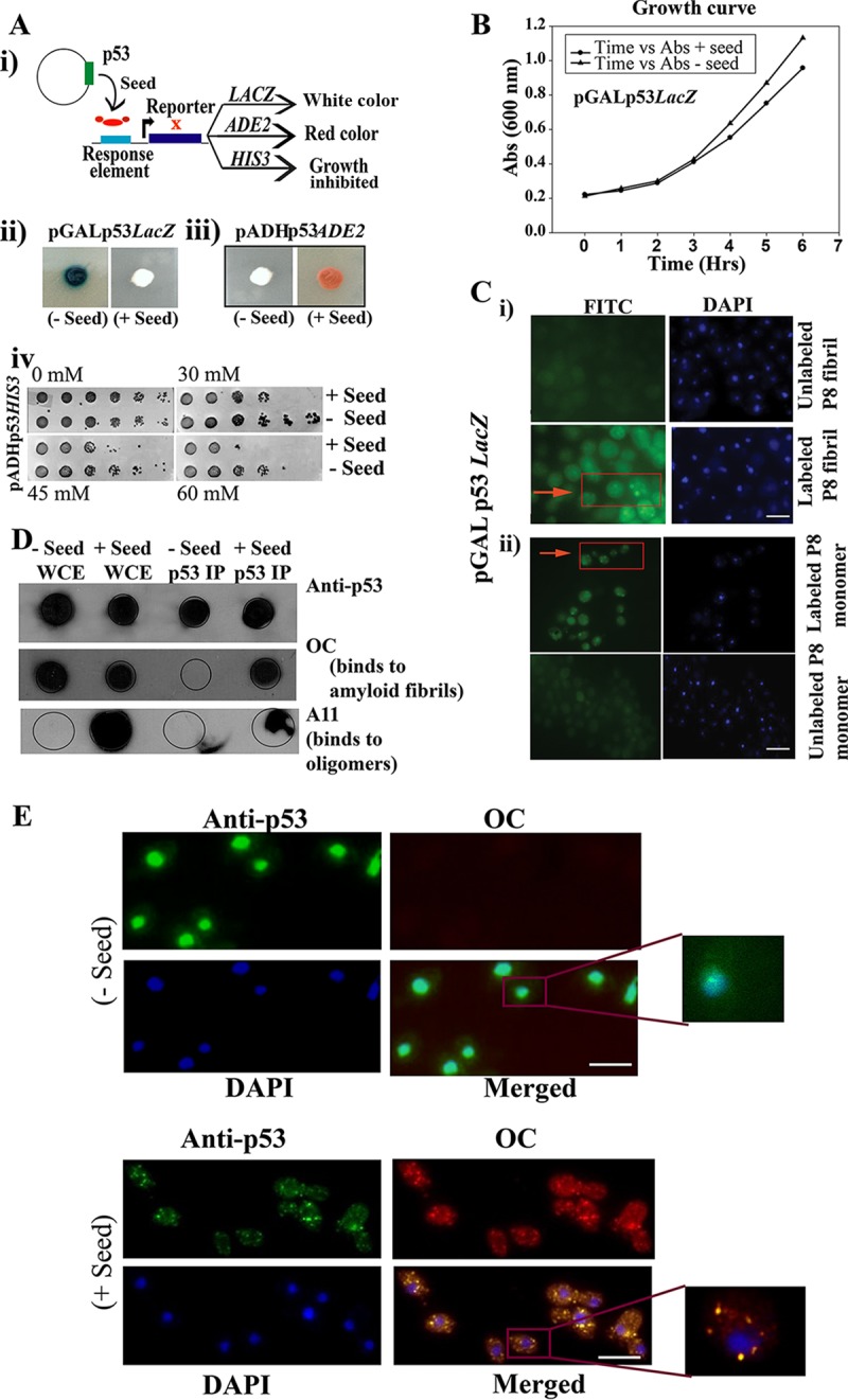
FIG 2.

Seeding effect of P8 fibrils (residues 250 to 257; PILTIITL) on functional p53. (A) Schematic representation of the loss of function of native p53 upon interaction with preformed P8 fibrils (Seed) (i), which leads to change in colony color (ii and iii) and a growth defect (iv), which is due to inactivation of transcription of three reporters, as indicated. (B) Growth curves of strain SGY6003 with (+) and without (−) seed were not found to be significantly different. (C) Internalization of FITC-labeled aggregated P8 fibrils and P8 monomer within the yeast cell. (i) FITC-labeled P8 fibril (green fluorescence) was used to transform yeast cells, similar to what was performed in panels A and E, and we obtained visual confirmation of the internalization of P8 fibrils and their localization in the cytoplasm. (Bottom left) Magnified image of the labeled seed (arrow) inside the cytoplasm. Scale bar, ∼2 μm. (ii) FITC-labeled P8 monomer (green fluorescence) was used to transform yeast cells, similar to what was performed in panels A and E, and we obtained visual confirmation of the internalization of P8 monomer and its localization in the cytoplasm (arrow). Scale bar, ∼2 μm. (D) Immunoprecipitation of p53 from cells with and without seed was performed using anti-p53 antibody. Dot blot analysis was performed with immunoprecipitated p53 using anti-p53, OC, and A11 antibodies separately. WCE, whole-cell extract; p53 IP, immunoprecipitated p53. (E) Immunofluorescence study showing colocalization of p53 and OC antibody in yeast cells with or without seeds. Aggregates of p53 are seen as green cytoplasmic focus structures specifically in the cells harboring the seeds, which showed robust colocalized signal with OC antibody. Scale bar, ∼5 μm.
