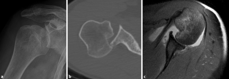
Fig. 3.
First-time posterior dislocation (A2) X‑ray (a) and computed tomography image (b) of an acute locked posterior shoulder dislocation with large reverse Hill–Sachs defect as well as magnetic resonance scan (c) of a reduced acute posterior shoulder dislocation with large Hill–Sachs defect and posterior Bankart lesion

