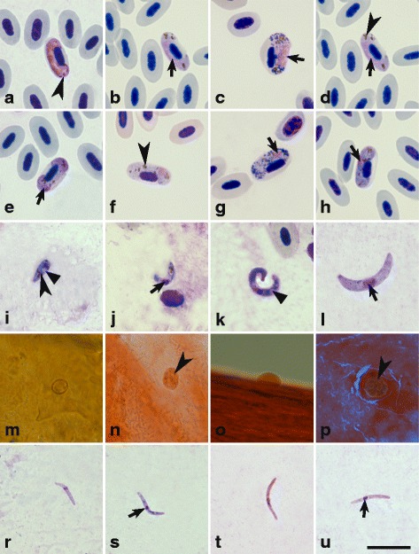
Fig. 1.

Mature microgametocytes (a-d), macrogametocytes (e-h), ookinetes (i-l), growing oocysts (m-p) and mature sporozoites (r-u) of Haemoproteus balmorali (c, g, k, o and t), H. majoris (b, f, j, n and s), H. motacillae (d, h, l, p and u) and H. pallidus (a, e, i, m and r). All images are from the methanol-fixed and Giemsa-stained thin films, except images m-p, which are from formalin-fixed whole mounts stained with Erlich’s haematoxylin. Short simple arrows: nuclei of parasites; arrowheads: pigment granules; triangle arrowheads: vacuoles. Scale-bar: 10 μm
