
Fig. 1.
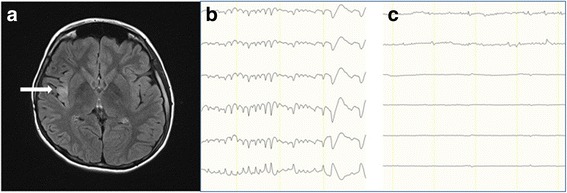
Insular hyperintensity shown on fluid-attenuated inversion-recovery MRI. 3-Tesla axial fluid-attenuated inversion-recovery images at the insular level show insular hyperintensity (a, arrow). The patient had an electrographic seizure on frontal strip of insular ECoG (b), which disappeared after insular resection (c)