
Fig. 1.
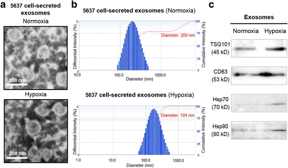
Characterization of normoxic and hypoxic exosomes derived from bladder cancer 5637 cells. a Transmission electron microscopy images of exosomes derived from normoxic and hypoxic 5637 cells. b Nanoparticle tracking analysis were analyzed the size distribution of normoxic and hypoxic exosomes derived from 5637 cells. c Western blotting analysis for exosomal markers TSG101, CD63, Hsp70 and Hsp90 of normoxic and hypoxic exosomes derived from 5637 cells; Equal amount of normoxic or hypoxic exosomes (500 ng) were used for western blotting analysis