
Fig. 3.
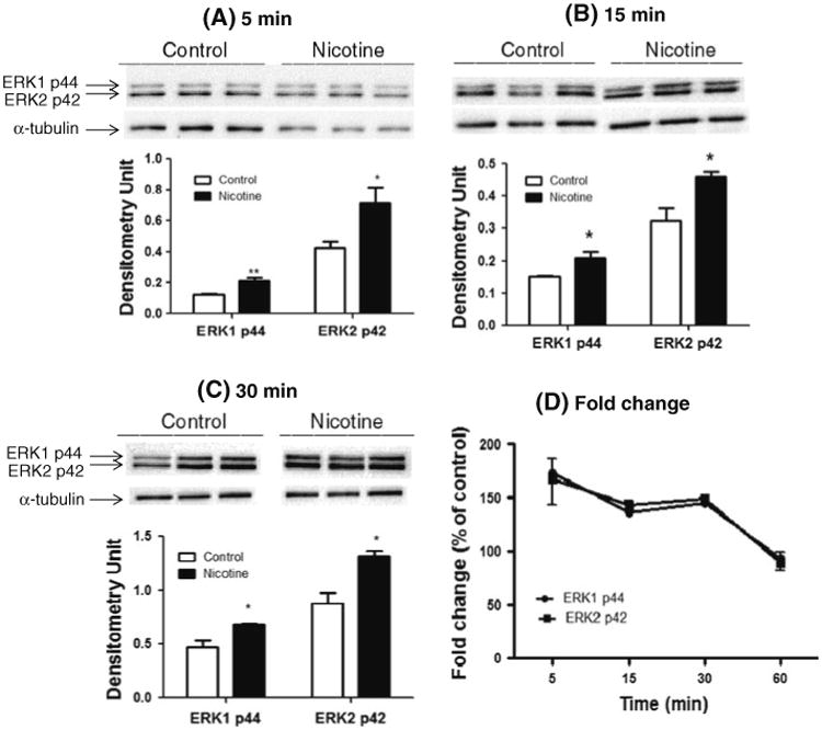
Activation of the ERK pathway by nicotine. Immunoblot analysis of whole-cell lysates from SH-SY5Y cells treated with nicotine for 5 min (a), 15 min (b), 30 min (c), and 1 h (data not shown) was performed to measure phosphorylation of ERK. Untreated cells were used as controls. In a, b and c, the upper panels are representative blots for Western analysis and the lower panel shows the mean ± SD of the optical density measurements (n = 3). The fold changes of p-ERK relative to untreated cells at each time point are shown in d as mean ± SD. **p < 0.01; *p < 0.05