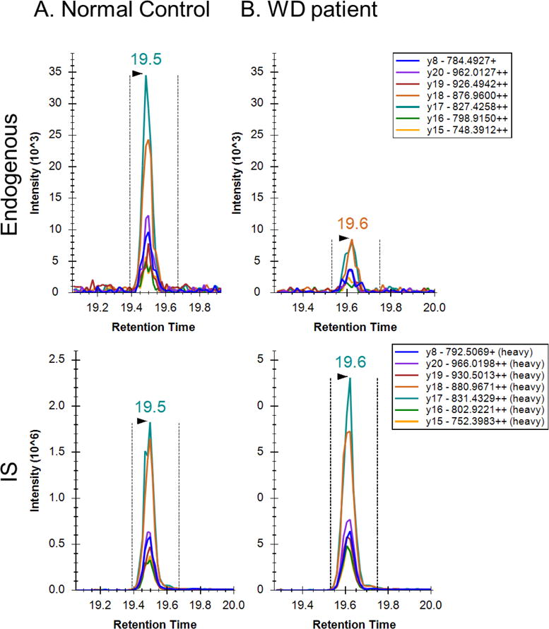
Figure 5. Extracted ion chromatograms for ATP7B 1056 peptide after peptide capture in DBS from (A) normal control and (B) WD patient.

Top panel is a signature peptide found in DBS. Bottom panel is the isotopically labeled internal standard. Chromatographic peaks overlap and SRM patterns are comparable. Transition labels refer to the precursor charge, fragment ion, fragment m/z, and fragment charge state.
