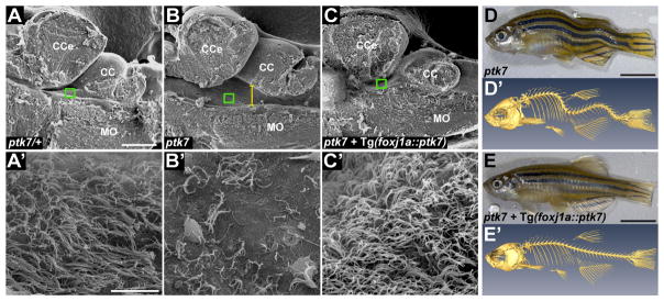
Fig. 1. ptk7 mutant fish exhibit hydrocephalus, EC cilia defects and spinal curves that are rescued by transgenic re-introduction of ptk7 specifically in motile ciliated cell lineages.
(A–C) Representative sagittal images of 2.5 month old ptk7/+ (A; n=6), ptk7 mutant (B; n=6) and ptk7 mutant expressing Tg(foxj1a::ptk7) (C; n=6) brains by SEM. Yellow line (B) demarcates hydrocephalus. Green squares depict corresponding high-magnification SEM images (A′–C′). (D–E′) Lateral views of fixed (D,E) and μCT rendered (D′,E′) representative adult ptk7 mutant (D and D′) and ptk7 mutant expressing Tg(foxj1a::ptk7) (E-E′). CCe - corpus cerebelli; CC - crista cerebellaris; MO - medulla oblongata. Scale bars: 250 μm (A,B,C); 10 μm (A′,B′,C′) and 5 mm (D,E).

