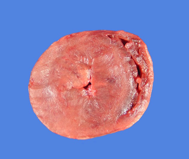
Figure 2.

A gross pathology specimen from a 5-year-old cat with hypertrophic cardiomyopathy. The heart is shown in a transverse plane at the level of the papillary muscles in the left ventricle. Severe concentric left ventricular hypertrophy is evident with dramatic reduction in the size of the left ventricular lumen.
