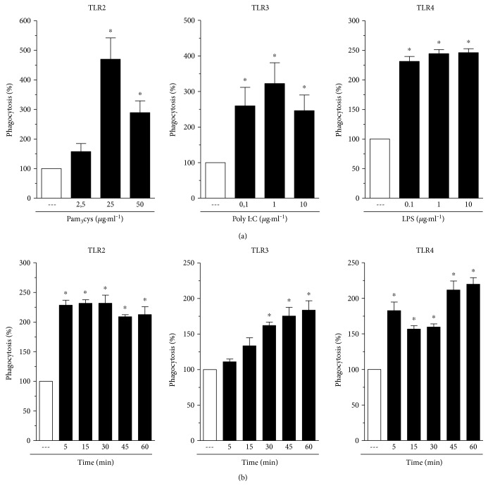
Figure 2.
TLR agonists increase IgG-sRBC phagocytosis in a dose- and time-dependent manner. (a) Rat AMs were cultured in the absence or in the presence of TLR2 (Pam3Cys), TLR3 (poly I:C), TLR4 (LPS) agonists at the indicated concentrations for 1 h. (b) The cells were cultured with TLR2 (Pam3Cys: 25 μg·ml−1), TLR3 (poly I:C: 1 μg·ml−1), or TLR4 (LPS: 1 μg·ml−1) at the indicated time periods. In both panels, rat AMs were challenged with RBCs or IgG-RBCs (1 : 40) after TLR agonist stimulation. Results are expressed as the mean ± SEM. Values are presented as the percentage of the IgG-RBC group. The RBC control group was discounted from all groups. ∗P < 0.05 compared to the IgG-RBC group. The experiment is a representative of three independent experiments performed in heptaplicate.

