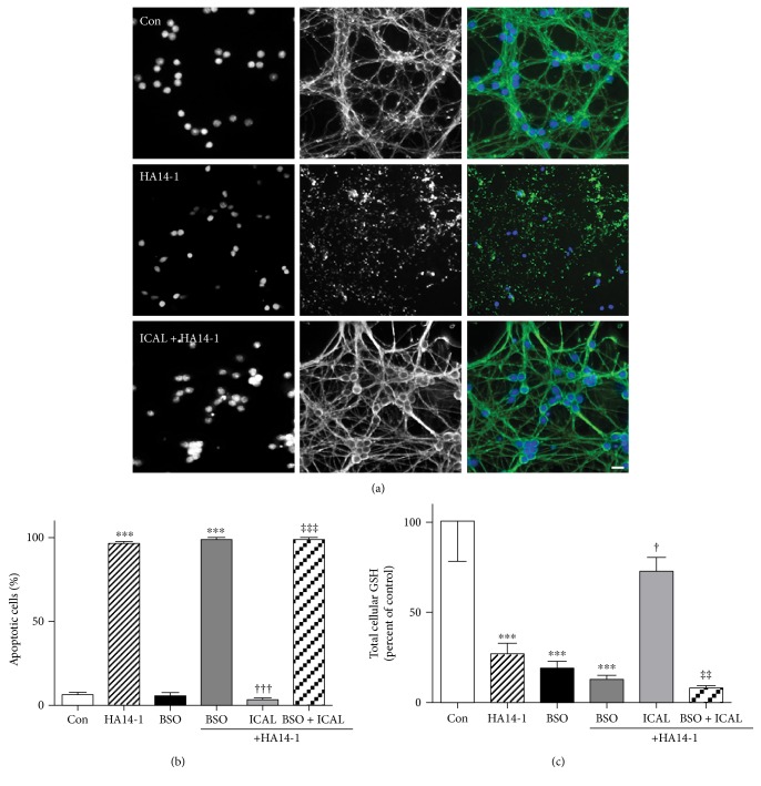
Figure 2.
Immunocal preserves cellular GSH and prevents apoptosis in CGNs exposed to the Bcl-2 inhibitor, HA14-1. (a) Representative images of CGNs left untreated (control), treated with HA14-1 (15 μM), or preincubated for 24 h with Immunocal before HA14-1 treatment for further 24 h. Panels from left to right, DAPI (nuclei), β-tubulin, merged images showing β-tubulin (green), and DAPI (blue). Scale bar, 10 μm. (b) Quantification of apoptosis for 4 independent experiments performed as in (a) except some cultures were preincubated with 200 μM BSO as well. Apoptotic cells were those with condensed or fragmented nuclei. Results are shown as mean ± SEM, n = 4. ∗∗∗ indicates p < 0.001 compared to control, ††† indicates p < 0.001 compared to HA14-1, ‡‡‡ indicates p < 0.001 compared to ICAL + HA14-1. (c) CGNs were treated exactly as described in (b). Total cellular GSH was measured as described in Materials and Methods. Data shown represent the percent of control cellular GSH concentration, mean ± SEM, n = 4. ∗∗∗ indicates p < 0.001 compared to control, † indicates p < 0.05 compared to HA14-1, and ‡‡ indicates p < 0.01 compared to ICAL + HA14-1. Significant differences were determined by one-way ANOVA with a post hoc Tukey's test. Con: control; ICAL: Immunocal; BSO: buthionine sulfoximine.

