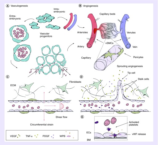
Figure 1. . Vascular network formation in vivo.
(A) Vascular progenitor cells directly form the dorsal aorta and cardinal vein (intra-embryonic) or coalesce into endothelial lined blood islands, which subsequently fuse into a primary plexus (extra-embryonic) and together form primary vasculature. Upon assembly, arteries and veins inosculate into smaller arterioles and venules (80–100 μm), and subsequent branching forms smaller capillaries and venules (10–15 μm). (B) Angiogenesis ensues and vessels mature and remodel, directed by a variety of angiogenic factors and cytokines (acidic FGF, bFGF, TGF-α), TFG-β, HGF, TNF-α, angiogenin, IL-8 and the angiopoietins [3]). (C) Pressure drives the perfusion of blood through capillaries, resulting in an efficient method for exchange of O2 and small molecules across the endothelium, and larger molecules, such as drugs, delivered through the endothelium. Connection to postcapillary venules drives the inward transport of CO2 and waste. Mechanical factors such as wall shear stress and axial strain also direct angiogenesis. (D) EC-secreted factors, such as TGF-β1, recruit mural cells during angiogenic remodeling. Pro-angiogenic factors are released from stromal cells such as fibroblasts, directing migration and sprouting of ECs. (E) Along with mural cells, and a continuously remodeling BM, ECs largely comprise the endothelium. Specialized organelles, WPBs, contained within ECs respond to various agonists (thrombin, histamine, VEGF, epinephrine, among others) by secretion of a variety of factors involved in wound healing and thrombosis. The most abundantly secreted glycoprotein is vWF. Secretion of vWF into circulating blood results in its binding to a blood-clotting factor, VIII, in its inactivated state. In the presence of thrombin, VIII is released, and vWF binds to glycoprotein Ib allowing for platelet adhesion and plug formation in sites of vascular injury.
BM: Basement membrane; EC: Endothelial cell; WPB: Weibel Palade bodies; vSMC: Vascular smooth muscle cell; vWF: von Willebrand Factor.
Concepts for Figure 1A & B are adapted with permission from [2].

