Figure 5.
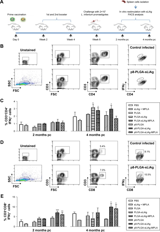
IFNγ-producing CD4+ and CD8+ T cells in vaccinated–infected BALB/c mice. (A) Groups of mice (n=5) were vaccinated with PBS, sLiAg + MPLA, PLGA, PLGA-sLiAg, PLGA-sLiAg-MPLA, p8-PLGA, p8-PLGA-sLiAg or p8-PLGA-sLiAg-MPLA three times with 2-week intervals. Two weeks after the second booster vaccination, mice were challenged with 2×10⁷ stationary-phase promastigotes of Leishmania infantum. Two and four months post-challenge, spleen cells were harvested and stimulated in vitro with sLiAg (12.5 μg/mL) for 72 h in 5% CO2 at 37°C. CD3+CD4+IFNγ+ and CD3+CD8+IFNγ+ cells were determined using flow cytometry. (B) Representative dot plots and (C) diagram showing IFNγ-producing CD4+ T cells 2 and 4 months post-infection. (D) Representative dot plots and (E) diagram showing IFNγ-producing CD8+ T cells 2 and 4 months post-infection. The results are the mean value ± SD of each group of mice. Significant differences between vaccinated and PBS control group are indicated by asterisks: *P<0.05, **P<0.01 or ***P<0.001.
Abbreviations: L. infantum, Leishmania infantum; PBS, phosphate-buffered saline; sLiAg, soluble Leishmania infantum antigens; MPLA, monophosphoryl lipid A; PLGA, poly(D,L-lactide-co-glycolide); p8, eight-amino-acid peptide; pc, post-challenge; FACS, fluorescence activated cell sorting; SSC, side-scattered light; FSC, forward-scattered light.

