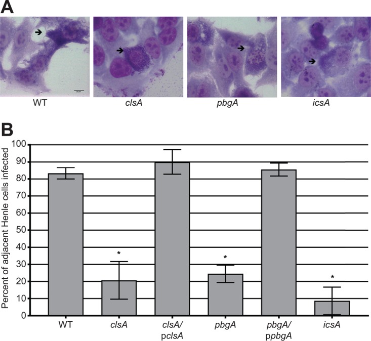
FIG 4 .

Cardiolipin is required for S. flexneri intercellular spread. Semiconfluent Henle monolayers were infected with approximately 107 CFU of bacteria. Monolayers were stained after 4 h, and intercellular spread was visualized by bright-field microscopy. (A) Micrographs of intercellular spread by WT S. flexneri and clsA, pbgA, and icsA mutants. Black arrows point to primary infected Henle cells. (B) Graphical representation of S. flexneri intercellular spread. One hundred infected Henle cells were counted positive for spread if the surrounding Henle cells were also infected. Values are means ± standard deviations (error bars) for three biological replicates. Values that are significantly different (P < 0.05) from the value for the wild type by Student’s t test are indicated by an asterisk.
