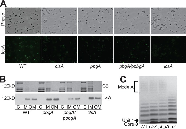
FIG 7 .
pbgA is required for unipolar IcsA localization. (A) Visualization of IcsA localization. Bacterial cultures were grown to mid-log phase (visualized by phase contrast microscopy), and IcsA was observed by indirect immunofluorescence (visualized by FITC). All images were captured with an exposure time of 1.5 s and processed in an identical manner. Bar = 5 μm. (B) Outer membrane IcsA levels. Bacteria were grown to mid-log phase. The membranes were fractionated using Sarkosyl membrane solubilization, resolved by (10%) SDS-PAGE, stained with Coomassie blue (CB) (same gel picture in Fig. 2B), and immunoblotted using either polyclonal anti-IcsA antisera. C, cytoplasm; IM, inner membrane; OM, outer membrane. (C) LPS structure of clsA and pbgA mutants. Bacteria were grown to mid-log phase. LPS was extracted, resolved by (4 to 12%) SDS-PAGE, and visualized by silver staining.

