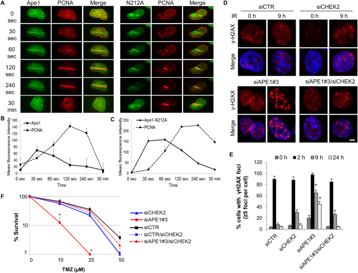
Figure 2.
Ape1 depletion results in DNA damage accumulation, and that is counteracted by concomitant depletion of Chk2 in glioblastoma cells. (A) U251-MG cells were exposed to laser micro-irradiation, and green (Ape1) and red (PCNA) fluorescence were live-imaged for 30 min. At least three independent irradiations were performed for each cell type. Representative images are shown. (B) Time-dependent damage site recruitments of Ape1, (C) Ape1-N212 A, and PCNA were determined as described in Methods. (D) U251-MG cells were exposed to IR (3 Gy) at 48 h of siRNA transfection. Cells were fixed at the indicated time points, and immunostained for γH2AX. Representative images are shown. Scale bar, 5 µm. (E) At least 100 cells per sample were counted from each experiment, and cells showing more than five γH2AX foci were quantified. Error bars indicate standard deviations obtained from three independent experiments. *p < 0.01 (siAPE1#3 vs. siAPE1#3/siCHEK2); Student’s t test. (F) U251-MG cells were transfected with the indicated siRNAs, and colony formation assay was performed after TMZ exposure. Data were obtained from three independent experiments. Error bars indicate standard deviation. *p < 0.05 (siAPE1#3 vs. siAPE1#3/siCHEK2); Student’s t test.

