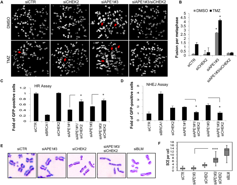
Figure 3.
Dual suppression of Ape1 and Chk2 facilitates HR. (A) U251-MG cells were transfected with the indicated siRNAs, and treated with TMZ (1000 µM, 1 h) at 48 h of transfection. Metaphase chromosomes were prepared 24 h after exposure to TMZ, and labelled with DAPI. Representative metaphases are shown. Red arrows indicate chromosome fusions. (B) At least 50 metaphases per sample were counted, and standard deviations were calculated from three independent experiments. *p < 0.001 (siAPE1#3 vs. siAPE1#3/siCHEK2); Student’s t test. (C) U2OS-DR cells were transfected with the indicated siRNAs and a plasmid expressing I-Sce I. Percentage of GFP-positive cells was quantified by flow cytometry. Percentage of values scored in the control cells (siCTR) was normalized to 1. Data were obtained from three independent experiments. *p < 0.05; Student’s t test. (D) H1299dA3-1 cells were transfected with the indicated siRNAs and an I-Sce I vector. Percentage of GFP-positive cells was calculated as described in (C). Data from three independent experiments are shown. *p < 0.05; Student’s t test. (E) U2OS cells were transfected with the indicated siRNAs, and incubated with BrdU (10 µM) for 24 h. After the treatment with MMS (350 µM) for 3 h, metaphase chromosomes were stained with Giemsa. Representative images are shown. (F) At least thirty metaphase cells per sample were analyzed, and medians of the number of chromosomes with SCE were presented in a box plot chart. ***p < 0.0001; (siAPE1#3 vs. siAPE1#3/siCHEK2); Student’s t test.

