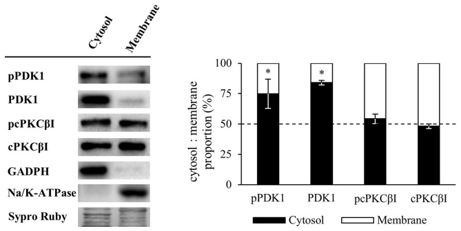
Figure 4.
Membrane and cytosol distribution of PDK1 and cPKCβI in basal conditions. Western Blot analysis of the distribution of PDK1 and cPKCβI in membrane and cytosol fraction of skeletal muscle. Results showed that in basal conditions, pPDK1 and PDK1 were found predominantly in the cytosol fraction while pcPKCβI and cPKCβI were present similarly in both cytosol and membrane fractions. Moreover, glyceraldehyde 3-phosphate dehydrogenase (GAPDH) was found in the cytosol fraction and essentially undetectable in the membrane fraction. As expected, the membrane protein Na/K-ATPase was highly enriched in this cellular component, and undetectable in the cytosol fraction. Data are mean percentage ± SEM, *p < 0.05 (n = 5). Abbreviations: phosphoinositide-dependent kinase 1; pPDK1, phosphorylated phosphoinositide-dependent kinase 1; cPKCβI, conventional protein kinase C βI; pPKCβI, phosphorylated conventional protein kinase C βI.

