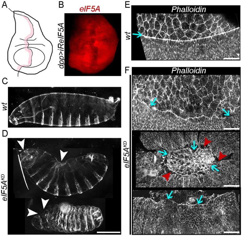
Figure 1.
Knockdown of Dm eIF5A in the embryonic epidermis produces defects in DC and actin cable assembly. (A) Schematic representation of the anterior-posterior (AP) compartment boundary of a Drosophila wing imaginal disc (shaded in pink). (B) Anti-eIF5A antibody staining of a dpp-GAL4/UAS-iReIF5A wing imaginal disc, in which eIF5A knockdown is induced in the AP boundary. (C,D) Dark-field micrographs of representative (C) wild-type and (D) eIF5A mutant embryos (eIF5A KD). Anterior is to the left, dorsal is up. White arrowheads point to holes or not properly sealed areas in the anterior and dorsal parts of the embryo. (E,F) Confocal images of the lateral epidermis, DME cells and AS cells of representative stage 13–14 (E) control and (F) eIF5A KD embryos stained with phalloidin. Blue arrows point to the actomyosin cable in DME cells, which is irregular and discontinuous in eIF5A mutants. Detachment of DME cells from AS cells is frequently observed in mutant embryos (red arrowheads in F). Scale bars: 100 µm in C,D; 25 µm in E,F.

