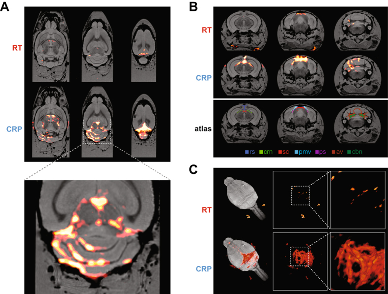
Figure 5.
High spatial resolution 19F MR image of an ex vivo brain from an EAE mouse showing clinical disease. With both 19 F-CRP and 19 F/1 H RT-coil, 19F MR images were acquired using a 3D-RARE sequence. 19F MR images (shown in red) were combined with 1H MR images (shown in grayscale). 1H MR images were acquired using a 3D-FLASH sequence and the 19 F/1 H RT-coil. (A) Three exemplary slices from horizontal views of combined 19F/1H MR images for both 19 F/1 H RT-coil (upper panel) and 19 F-CRP (middle panel), in the lower panel a 300% zoom of the 19F/1H MR images acquired with the CRP. (B) Three exemplary slices from coronal views of combined 19F/1H MR images for both RT coil (upper panel) and CRP (middle panel). Registration of the Allen brain atlas to the 1H image (lower panel) shows following labelled brain regions: rs: retrosplenial area; crn: cranial nerves; sc: superior colliculus (sensory related); pmv: posteromedial visual area; ps: postsubiculum; av: arbor vitae; cbn: cerebellar nuclei. (C) 3-D rendering of the combined 19F/1H MR images for both 19 F/1 H RT-coil (upper panel) and 19 F-CRP (lower panel).

