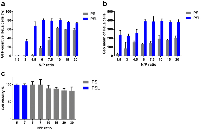
Figure 1.
Transfection efficiency and cytotoxicity of PSL and PS complexes in HeLa cells at various N/P ratios in the presence of 10% FCS. Transfection was performed at a dose of 0.3 μg of DNA per well in a total volume of 200 μl. GFP expression was quantified by flow cytometry after 24 hours. (a) Percentage of GFP-positive HeLa cells. (b) Geometric mean of fluorescence intensity of GFP-positive cells. (c) Cell viability assay of HeLa cells after transfection. Data represent mean ± standard deviation of 3 independent experiments.

