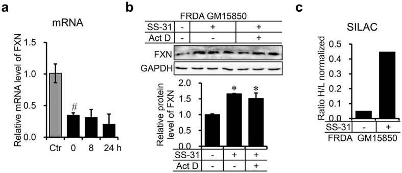
Figure 6.
SS-31 treatment translationally upregulates FXN expression. (a) mRNA level of FXN in GM15850 cells after treatment with 50 nM of SS-31 for 8 or 24 h. Total RNA was extracted from the cells after treatment and analysed by qRT-PCR. The relative level was normalised to that in healthy GM15849 cells. GAPDH was used as an internal control. Values represent mean ± SEM (n = 4, each duplicates). (b) Protein level of FXN in GM15850 cells after co-treatment with 50 nM of SS-31 and 0.5 µg/mL of actinomycin D (ActD). The cells were collected after 8 h and the FXN protein level was detected by Western blotting. Upper panel: a representative graph of western results; lower panel: quantitative data of the intensity of the western bands. Values represent mean ± SEM (n = 3, each duplicates). A one-way ANOVA was carried out, # p < 0.05 compared with the healthy control GM14519 cells. *p < 0.05 compared with the untreated GM15850 cells. (c) Ratio of newly synthesised FXN after SS-31 treatment to before-treatment synthesised protein revealed by SILAC technology in GM15850 cells. The Cells were cultured in conventional RPMI 1640 medium containing 12C14N-Lysine and 12C14N-Arginine (“light”, L) overnight, then transferred into medium containing 13C6 15N2-Lysine and 13C6 15N4-Arginine (“heavy”, H) for 24 h with/without 50 nM of SS-31 (see Materials and Methods). Immunoprecipitation was performed with FXN antibody to enrich total FXN protein, which was sent for SILAC assay. No significance data for (c) due to only two repeats.

